Definition and Pathophysiology
Osteoarthritis is defined as a disorder of synovial joints which occurs when damage triggers repair processes leading to structural changes within a joint.
These repair processes alter the structure of the joint over time, causing typical features of:
- Localised loss of cartilage.
- Remodelling of adjacent bone and the formation of osteophytes (new bone at joint margins).
- Mild synovitis (inflammation of the synovial membrane that lines the joint capsule).
In some people, these repair processes may alleviate symptoms, but in others they cannot fully compensate for the joint damage, and symptoms of pain and stiffness may occur.
Joint damage may occur through repeated excessive loading and stress of a joint over time, or by injury. Any synovial joint can be involved, and the most commonly affected peripheral joints are the knees, then the hips, and the small joints of the hand.
Risk Factors
The exact causes of osteoarthritis are not fully known. It is a complex and multifactorial condition involving genetic, biological, and biomechanical components.
Genetics
- Family history of osteoarthritis
- Genetic predisposition to joint degeneration
Biological
- Increasing age
- Female sex
- Obesity
- High or low bone density
Biomechanical
- Joint injury and damage
- Occupational and recreational stresses on joints
- Joint laxity and reduced muscle strength
- Joint malalignment
Clinical Features
Suspect a diagnosis of osteoarthritis if alternative conditions have been excluded and a person is aged 45 years or more, with suggestive clinical features.
Symptoms
- Activity-related joint pain — typically only one or a few joints are affected at any one time, and pain develops over months or years
- No morning joint-related stiffness, or morning stiffness lasting no longer than 30 minutes
Signs
- Bony swellings and joint deformities
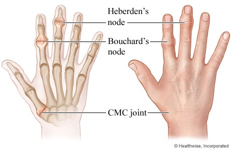
- Bouchard's nodes (bony nodules on proximal interphalangeal joints)
- Heberden's nodes (bony nodules on distal interphalangeal joints)
- Joint effusions
- Joint warmth and/or tenderness
- Muscle wasting and weakness
- Restricted and painful range of joint movement
- Joint crepitus
- Joint instability
- Antalgic gait
Diagnostic Imaging
Routine X-ray of the affected joint(s) is not usually needed to confirm the diagnosis. Consider arranging an X-ray, depending on clinical judgement:
- If there is diagnostic uncertainty
- To exclude alternative conditions
- If there is a sudden clinical deterioration in symptoms
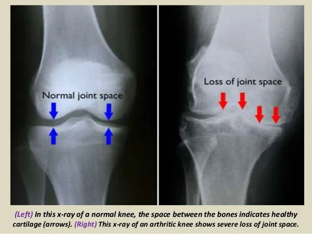
Typical radiological features of osteoarthritis include:
- Subchondral bone thickening (sclerosis) and/or cysts
- Osteophyte formation (new bone formation at joint margins)
- Loss or narrowing of the joint space (provides an estimate of the severity of cartilage damage)
Note: structural changes on X-ray may not correlate with reported symptoms and functional impairment.
Management
Analgesia
- Paracetamol
- Topical NSAIDs
- Oral NSAIDs
- Opioids
- Topical capsaicin
- Intra-articular corticosteroids for moderate-to-severe joint pain
Physical treatments
- Strengthening exercises
- Aerobic fitness training
- Physiotherapy
- Assisted devices for activities of daily living
- Local heat/cold packs
- Supports and braces for biomechanical joint pain/instability
- Appropriate footwear for lower limb arthritis
Surgery
- Refer if the person has joint symptoms (pain, stiffness, and reduced function) that have a substantial impact on their quality of life and are refractory to non-surgical treatment
- Surgical treatment usually involves joint replacement or fusion. Arthroscopic lavage and debridement may be considered for people with knee osteoarthritis with a clear history of mechanical locking. Hand involvement may be treated by excision of the trapezium or joint fusion
- Complications of joint replacement may include aseptic loosening, pain, dislocation, infection, and fracture
Complications
- Joint deformity
- Functional impairment and disability
- Psychosocial impact
- Occupational impact
- Falls
- Chronic pain syndrome