Pulmonary Embolism (PE)
Obstruction of pulmonary arteries by emboli, typically from venous thrombi
Definitions
Pulmonary embolism (PE) is a condition in which one or more emboli, usually arising from a thrombus formed in the veins (or, rarely, in the right heart), are lodged in and obstruct the pulmonary arterial system, causing severe respiratory dysfunction.
Provoked PE
- Associated with antecedent (within 3 months) transient risk factors
- Examples: immobility, surgery, trauma, pregnancy/puerperium, OCP/HRT use
- Risk factors can be removed → lower recurrence risk
Unprovoked PE
- Occurs without transient risk factors
- May have persistent risk factors (cancer, thrombophilia)
- Higher recurrence risk as factors can't be removed
Pathophysiology
Key Mechanisms
- Ventilation-perfusion mismatch → intrapulmonary dead space
- Reduced pulmonary arterial cross-section → ↑pulmonary pressure, ↓cardiac output
- Alveolar collapse after hours → worsens hypoxemia
- Large/multiple emboli → acute right ventricular failure
Chronic Thromboembolic PH (CTEPH)
Rare complication where emboli become fibrous tissue → chronic obstruction → progressive pulmonary hypertension → right heart failure
Risk factors: inadequate anticoagulation, large thrombus mass, residual thrombi, recurrent VTE
Sources of Emboli
- Deep vein thrombosis (most common)
- Tumors (prostate, breast)
- Fat (long-bone fractures)
- Amniotic fluid (pregnancy)
- Foreign bodies (IV drug use)
- Air (surgery)
- Sepsis (infective endocarditis)
Risk Factors
Major Risk Factors
- Current DVT
- Previous DVT/PE
- Active malignancy
- Recent surgery/hospitalization/immobilization
- Pregnancy and up to 6 weeks postpartum
Other Risk Factors
- Age >60 years
- OCP/HRT use
- Obesity (BMI >30)
- Long-distance sedentary travel
- Varicose veins
- Thrombophilias
- Medical comorbidities
Virchow's Triad
30-50% of VTE episodes have no identifiable risk factor. Others result from:
- Hypercoagulability
- Venous stasis
- Vascular wall damage/dysfunction
Clinical Features
Symptoms
- Dyspnea (most common)
- Tachypnea
- Pleuritic chest pain
- Cough/hemoptysis
- Dizziness/syncope
- DVT symptoms
Signs
- Tachycardia
- Hypotension (massive PE)
- Hypoxemia
- Elevated JVP
- Gallop rhythm, split S2, TR murmur
- Pleural rub
- DVT signs
Investigations
Initial Workup
- ECG: Look for right heart strain (S1Q3T3, RBBB, T-wave inversion V1-V3)
- CXR: Mainly to exclude alternatives (may show atelectasis, effusion, elevated hemidiaphragm)
- ABG: If SpO2 <92% (hypoxia, increased A-a gradient)
Diagnostic Imaging
- CTPA: Investigation of choice for high probability PE or positive D-dimer
- V/Q scan: Alternative in pregnancy or normal CXR with no chronic disease
- Lower limb ultrasound: Useful in pregnancy to avoid irradiation
Echocardiography (for massive PE)
- Right ventricular dilation (>60% LV size) and hypokinesis
- McConnell's sign: RV free wall akinesis with apical sparing
- Paradoxical septal motion ("D-sign" on parasternal short-axis)
- IVC distension/non-collapsibility
- Tricuspid regurgitation
PE Wells Score
| Criteria | Points |
|---|---|
| Clinical signs/symptoms of DVT | 3 |
| Alternative diagnosis less likely than PE | 3 |
| Heart rate >100 bpm | 1.5 |
| Immobilization ≥3 days or surgery in previous 4 weeks | 1.5 |
| Previous PE/DVT | 1.5 |
| Hemoptysis | 1 |
| Active malignancy | 1 |
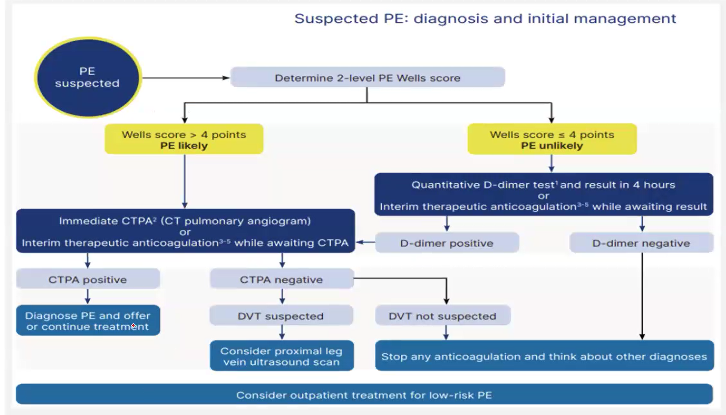
Wells ≤4 (PE unlikely)
- D-dimer test within 4 hours if possible
- If D-dimer delayed → interim anticoagulation
- Positive D-dimer → manage as Wells >4
- Negative D-dimer → stop anticoagulation, consider alternatives
Wells >4 (PE likely)
- CTPA immediately if possible
- If CTPA contraindicated → V/Q SPECT or planar scan
- If imaging delayed → interim anticoagulation
- Confirmed PE → continue anticoagulation
- No PE → stop anticoagulation, consider alternatives
- DVT suspected: Consider proximal leg vein ultrasound scan
- DVT not suspected: Stop interim therapeutic anticoagulation (except for COVID-19)
- Consider outpatient treatment for low-risk PE
Management
Anticoagulation
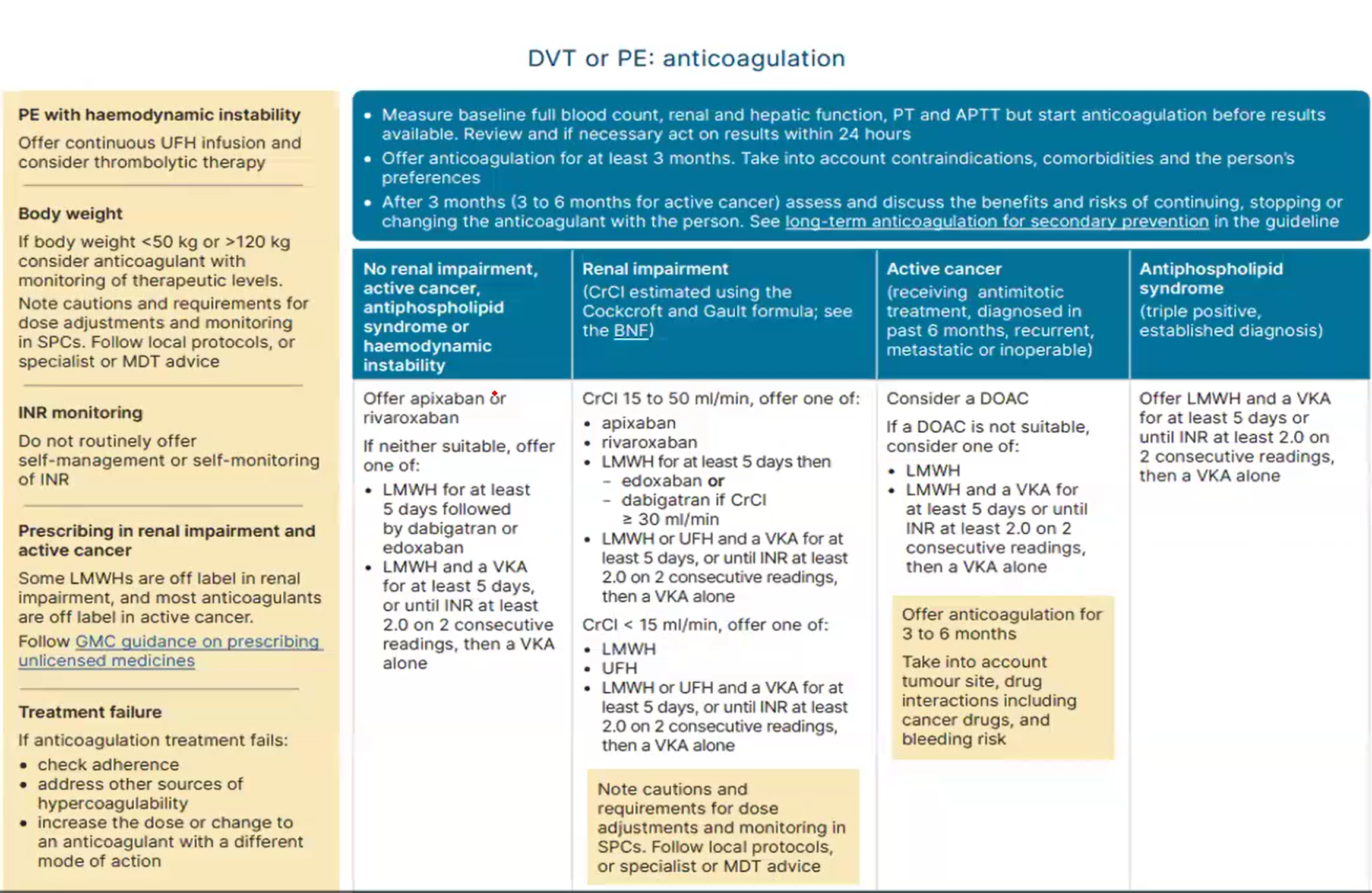
- Baseline bloods (FBC, renal/liver function, PT/APTT) - don't delay treatment
- First line: Apixaban or rivaroxaban unless contraindicated
- Alternatives:
- LMWH for ≥5 days → dabigatran/edoxaban OR
- LMWH + VKA until INR ≥2.0 for 2 readings → VKA alone
- Avoid UFH unless renal impairment/bleeding risk
- Minimum 3 months treatment for confirmed PE
Thrombolytic Therapy
- Consider for PE with hemodynamic instability
- Do not use for stable PE (with/without RV dysfunction)
Mechanical Interventions
- IVC filters: Trap emboli while preserving blood flow
- Indications:
- Contraindication to anticoagulation
- Recurrent PE despite adequate anticoagulation
- May be temporary or permanent
Anticoagulation in Special Populations
| Condition | CrCl Range | Treatment Options | Notes |
|---|---|---|---|
| Renal Impairment | CrCl 15-50 mL/min |
1st line: Apixaban or Rivaroxaban If not suitable: Other options |
|
| CrCl <15 mL/min | Avoid DOACs in severe renal impairment | ||
| Active Cancer | Any | LMWH is preferred over DOACs for most cancer-associated thrombosis | |
| Antiphospholipid Syndrome | Any | Warfarin is preferred (target INR 2-3 or 3-4 depending on risk) | |
| None of the above | Any | ||
Important Notes:
- For LMWH + warfarin or UFH + warfarin: Continue for at least 5 days or until INR ≥2.0 on 2 consecutive readings, then warfarin alone
- Monitor renal function regularly during treatment
- Consider bleeding risk when selecting anticoagulant
- Dose adjustments may be required for renal impairment
Clinical Images