Spontaneous Pneumothorax
Air in the pleural space without trauma or medical intervention
Definitions
Primary Spontaneous Pneumothorax (PSP)
- Occurs without suspected lung disease
- Most have emphysema-like changes on CT
- Different management and outcomes from SSP
Secondary Spontaneous Pneumothorax (SSP)
- Occurs with underlying lung disease
- Consider secondary if >50 years with smoking history
- Associated with:
- COPD (most common)
- TB, asthma, pneumonia
- Sarcoidosis, cystic fibrosis
- Malignancy, bronchiectasis
- Pulmonary fibrosis, AIDS
Risk Factors
Primary Spontaneous Pneumothorax
- Tall, thin males
- Marfan's syndrome
- Ehlers-Danlos syndrome
- Alpha-1 antitrypsin deficiency
- Smoking
Clinical Features
Symptoms
- Dyspnea
- Tachypnea
- Chest pain
Signs
- Reduced expansion on affected side
- Reduced air entry
- Hyperresonance to percussion
- Reduced vocal/tactile fremitus
Key Differences
- PSP: Symptoms mild, often present >2 days after onset
- SSP: More severe symptoms due to pre-existing lung disease
- Tension pneumothorax: Consider if severe symptoms + cardiorespiratory distress
Investigations
Chest X-ray
- Erect PA view in inspiration
- Findings:
- Visible visceral pleural edge (thin white line)
- Absent lung markings peripherally
- Radiolucent peripheral space
- Small pleural effusions sometimes seen
CT Scanning
- Gold standard for small pneumothoraces
- Useful for:
- Surgical emphysema
- Bullous lung disease
- Aberrant chest drain placement
- Additional lung pathology
Arterial Blood Gas
- Not needed if SpO2 >92% on room air
- 75% have PaO2 <10.9 kPa
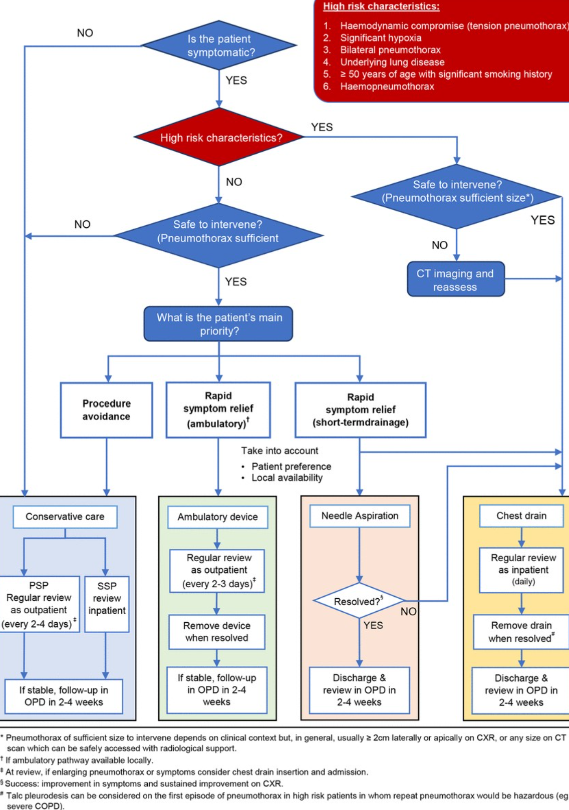
Management (BTS 2023 Pathway)
Initial Assessment
Is the patient symptomatic?
- No:
- Conservative care
- PSP: Outpatient review every 2-4 days
- SSP: Inpatient review
- If stable: OPD follow-up in 2-4 weeks
- Yes: Assess for high risk characteristics:
- Hemodynamic compromise (tension)
- Significant hypoxia
- Bilateral pneumothorax
- Underlying lung disease
- >50 years + smoking history
- Hemopneumothorax
High Risk Characteristics Present?
- Yes + unsafe to intervene: CT imaging and reassess
- Yes + safe to intervene:
- Chest drain
- Inpatient review
- Remove drain when resolved
- Consider talc pleurodesis for first episode in high-risk patients
- OPD follow-up in 2-4 weeks
- No + unsafe to intervene: Conservative care
- No + safe to intervene: Consider (patient preference/local availability):
- Conservative care OR
- Ambulatory device + outpatient review every 2-3 days OR
- Needle aspiration
- If not resolved → chest drain
Note: Intervention usually considered for ≥2cm laterally/apically on CXR or any size on CT that can be safely accessed
Discharge and Follow-up
Patient Advice
- Return if increasing breathlessness develops
- Respiratory specialist follow-up until full resolution
- Avoid air travel for ≥1 week after confirmed resolution
- Permanent diving restriction (unless bilateral surgical pleurectomy with normal post-op lung function/CT)
- Smoking cessation (reduces recurrence risk)