Introduction
Deep vein thrombosis (DVT) is the term used to describe the formation of a thrombus (blood clot) in a deep vein, which partially or completely obstructs blood flow. Thrombosis usually affects the deep veins of the legs or pelvis, but may affect other sites.
Complication Risk
The thrombus can dislodge and travel in the blood, especially to the pulmonary arteries. This is known as a pulmonary embolism (PE).
Classification
DVT may be classified as provoked or unprovoked based on the presence of transient risk factors.
Provoked DVT
DVT associated with a transient risk factor such as significant immobility, surgery, trauma, and pregnancy or puerperium. The combined contraceptive pill and hormone replacement therapy are also considered to be provoking risk factors.
Unprovoked DVT
DVT occurring in the absence of a transient risk factor. The person may have no identifiable risk factor or a risk factor that is persistent and not easily correctable (such as active cancer or thrombophilia).
Risk Factors
Risk Factors
Deep vein thrombosis (DVT) is more likely to occur in people with continuing or intrinsic risk factors.
Persistent Risk Factors
- A history of DVT
- Cancer (known or undiagnosed)
- Age over 60 years
- Being overweight or obese
- Male sex
- Heart failure
- Medical illness (e.g., acute infection)
- Acquired or familial thrombophilia
- Inflammatory disorders
- Varicose veins
- Smoking
Transient Risk Factors
- Recent major surgery
- Recent hospitalisation
- Recent trauma
- Chemotherapy
- Significant immobility
- Prolonged travel (for more than 4 hours)
- Significant trauma or direct trauma to a vein
- Hormone treatment
- Pregnancy and the postpartum period
- Dehydration
Clinical Features
Consider the possibility of deep vein thrombosis (DVT) if typical symptoms and signs are present, especially if the person has risk factors such as previous venous thromboembolism and immobility.
- Unilateral localised pain (usually throbbing) that occurs when walking or bearing weight
- Calf swelling (or more rarely, swelling of the entire leg)
- Tenderness
- Skin changes (edema, redness, and warmth)
- Vein distension
Differential Diagnosis
Several conditions can mimic DVT and should be considered in the differential diagnosis.
Physical Trauma
- Calf muscle tear/Achilles' tendon tear
- Calf muscle haematoma or fracture
Cardiovascular Disorders
- Superficial thrombophlebitis
- Post-thrombotic syndrome
- Venous obstruction or insufficiency
- External compression of major veins
- Arteriovenous fistula and congenital vascular abnormalities
- Acute arterial ischaemia
- Vasculitis
- Heart failure
Other Conditions
- Ruptured Baker's cyst
- Cellulitis
- Dependent (stasis) oedema
- Lymphatic obstruction
- Septic arthritis
- Cirrhosis
- Nephrotic syndrome
- Compartment syndrome
Management
Diagnostic Approach
Use the two-level DVT Wells score to assess the likelihood of DVT and inform further management.
Clinical Images
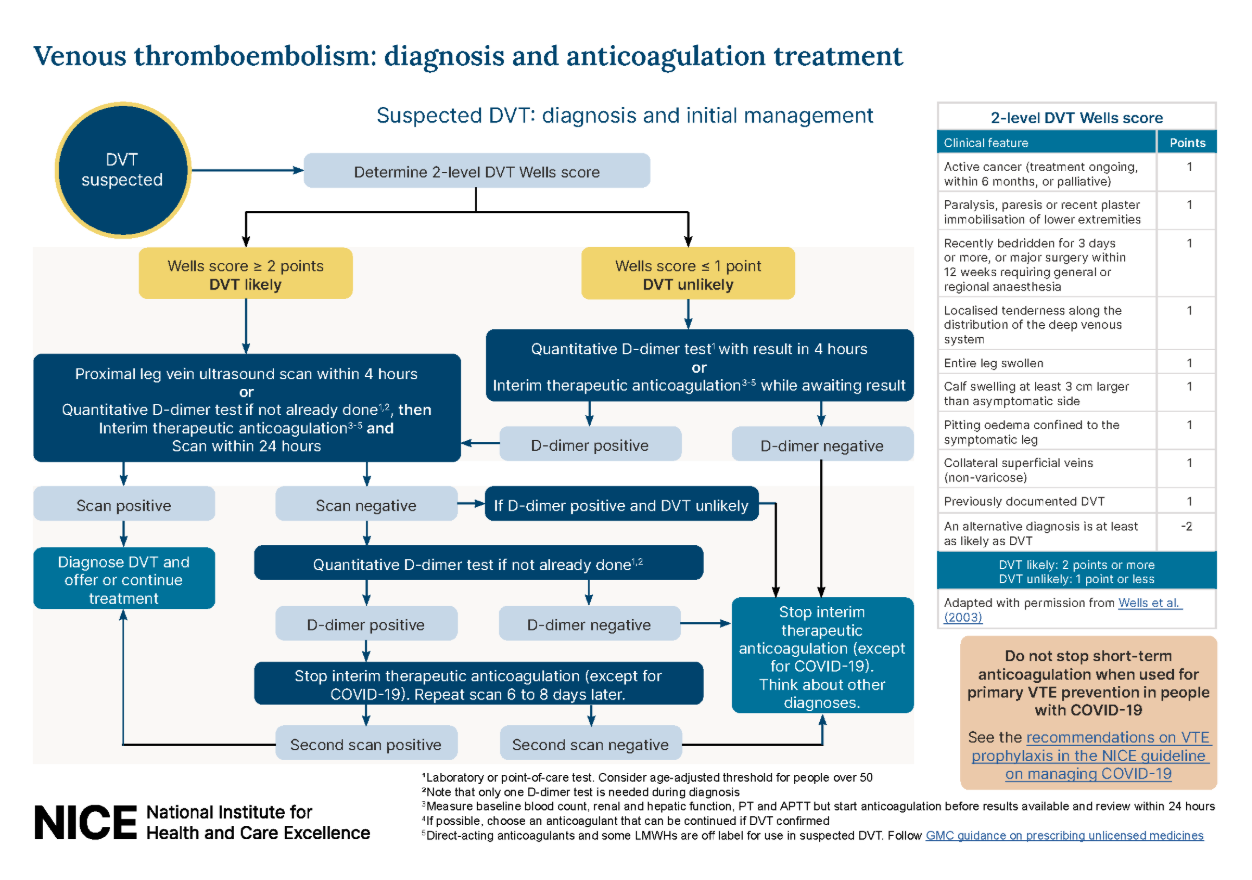
DVT Wells Score
- Active cancer (treatment ongoing, within the last 6 months, or palliative)
- Paralysis, paresis, or recent plaster immobilization of the legs
- Recently bedridden for 3 days or more, or major surgery within the last 12 weeks requiring general or regional anesthesia
- Localised tenderness along the distribution of the deep venous system
- Entire leg is swollen
- Calf swelling by more than 3 cm compared with the asymptomatic leg
- Pitting oedema confined to the symptomatic leg
- Collateral superficial veins (non-varicose)
- Previously documented DVT
Management Based on Wells Score
- Offer a proximal leg vein ultrasound scan with the results available within 4 hours if possible
- If ultrasound cannot be carried out within 4 hours, offer D-dimer test, interim therapeutic anticoagulation, and ultrasound within 24 hours
- Offer a D-dimer test with results available within 4 hours
- If D-dimer positive, offer ultrasound within 4 hours or interim anticoagulation and ultrasound within 24 hours
- If D-dimer negative, stop interim anticoagulation and consider alternative diagnosis
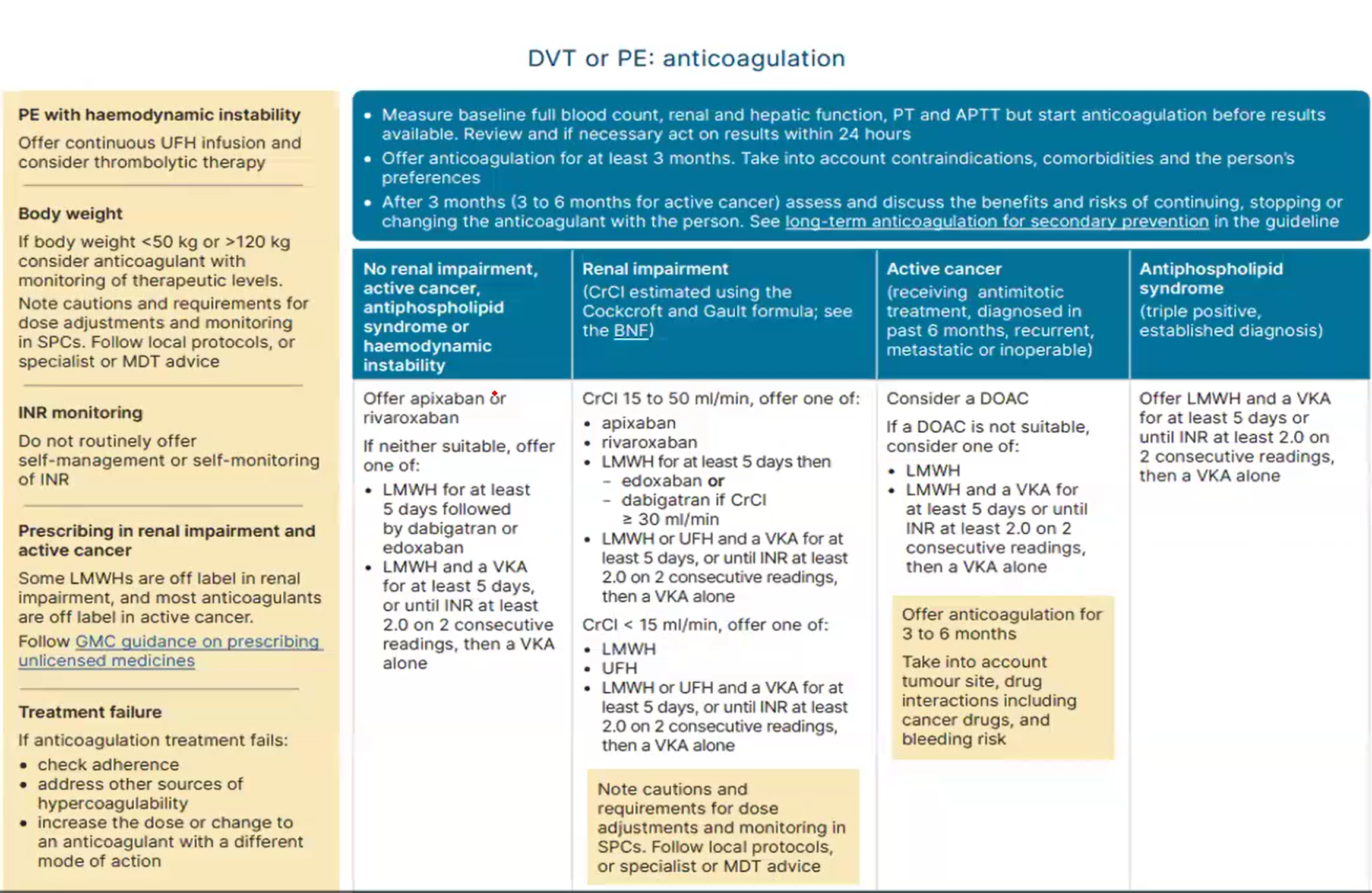
- Offer apixaban or rivaroxaban first line
- Alternative options: LMWH for at least 5 days followed by dabigatran or edoxaban
- Carry out baseline blood tests but do not wait for results before starting treatment
Follow-up of Confirmed DVT
Complications
Serious Complications
The most serious complication of deep vein thrombosis (DVT) is death due to pulmonary embolism (PE).